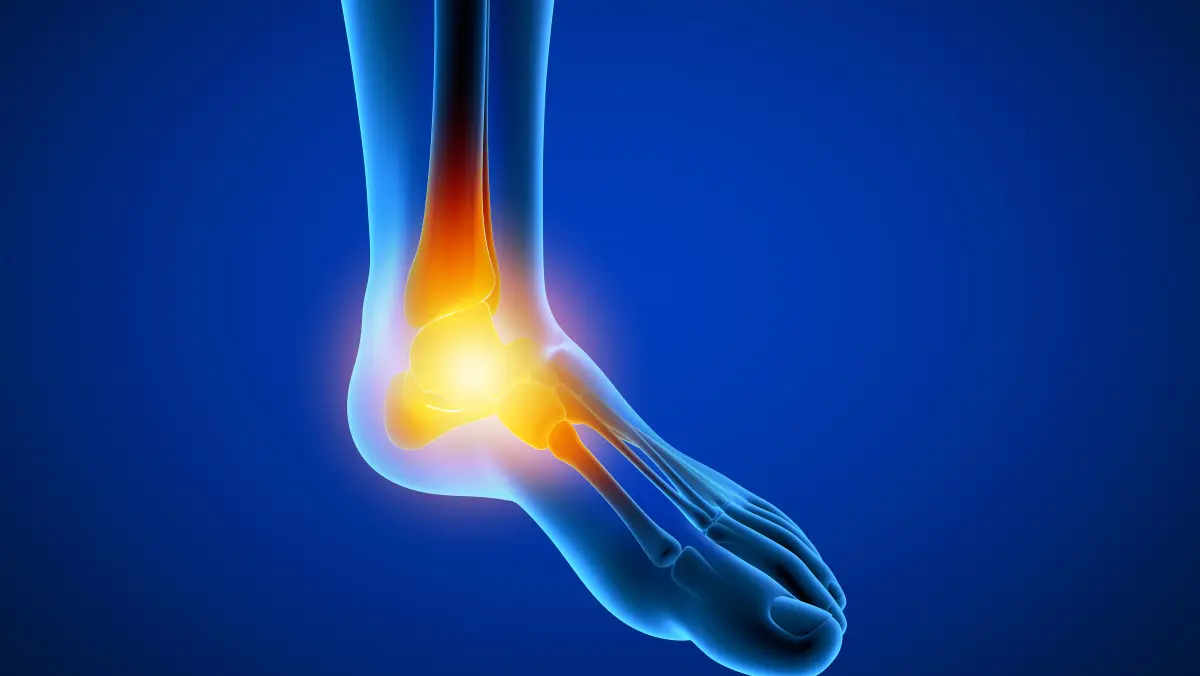
Os trigonum syndrome is a foot condition that results from the formation of a small, additional bone called the os trigonum. This bone is present in about 7-15% of the general population and is located at the back of the ankle, behind the ankle bone (lateral malleolus). When the os trigonum causes pain or impingement, arthroscopic excision may be recommended to relieve the symptoms.
Arthroscopic excision of os trigonum impingement is a minimally invasive surgical procedure performed using an arthroscope, a small camera, and specialized instruments inserted through small incisions in the skin. The procedure involves removal of the os trigonum bone to relieve pain and improve mobility. It is typically performed as an outpatient procedure under local anesthesia with sedation or general anesthesia.
Following the procedure, the patient may experience some pain, swelling, and stiffness in the ankle joint. Rehabilitation is important to regain full range of motion and strength of the ankle. Physical therapy may be recommended to aid in recovery and prevent future injury. Full recovery may take several weeks to several months, depending on the individual case.
Arthroscopic excision of Os trigonum impingement is a minimally invasive surgical procedure performed to treat the pain and limited range of motion caused by an extra bone at the back of the ankle called the Os trigonum. This bone can cause impingement between the ankle and the back of the leg, leading to inflammation, pain, and stiffness. Arthroscopic excision of Os trigonum involves the removal of this extra bone using an arthroscope, which is a small camera attached to a tube inserted through a small incision in the ankle. The procedure is usually performed on an outpatient basis and can help relieve pain and improve ankle function. Rehabilitation after surgery typically involves a combination of rest, physical therapy, and gradual return to normal activities.
Preoperative considerations for arthroscopic excision of Os trigonum may include:
Medical evaluation: Before the surgery, your surgeon may conduct a thorough medical examination to assess your overall health status and any underlying medical conditions that may affect the surgery’s outcome.
Diagnostic tests: You may undergo imaging tests such as X-rays, MRI, or CT scans to confirm the presence of the os trigonum and the extent of the impingement.
Medications: Your surgeon may recommend stopping certain medications that can increase the risk of bleeding during and after surgery, such as aspirin, nonsteroidal anti-inflammatory drugs (NSAIDs), and blood-thinning medications.
Fasting: You may need to fast for a certain period before the surgery, typically around eight hours, to reduce the risk of complications during anesthesia.
Anesthesia: The procedure is usually performed under general anesthesia or regional anesthesia, such as a spinal or epidural block. Your anesthesia provider will discuss the options with you and help you choose the most appropriate type of anesthesia based on your health status and personal preferences.
Planning for recovery: You should make arrangements for a responsible adult to drive you home after the surgery and stay with you for the first 24 hours. You may also need to take some time off work and arrange for assistance with daily activities, such as cooking, cleaning, and bathing, during the recovery period.
Arthroscopic excision of Os trigonum is a minimally invasive surgical procedure that involves removing a small bone at the back of the ankle joint known as the os trigonum. This bone can sometimes cause pain and impingement in the ankle joint, especially in dancers, gymnasts, and athletes who frequently use their feet and ankles.
The arthroscopic excision of Os trigonum is typically performed under general anesthesia, and a small incision is made in the back of the ankle to insert an arthroscope and other surgical instruments. The surgeon then identifies and removes the os trigonum, which is usually done by carefully cutting and removing any soft tissues that may be attached to the bone.
After the os trigonum is removed, the surgical incision is closed, and a sterile dressing is applied. The patient is typically allowed to go home on the same day, but they will need to keep the affected leg elevated for a few days to help reduce swelling and pain.
Physical therapy is usually recommended after the procedure to help regain range of motion and strengthen the ankle joint. Most patients are able to return to their normal activities within a few weeks to a few months after the surgery, depending on the severity of their condition and their individual healing process.
After the arthroscopic excision of Os triennium, the patient will be moved to the recovery room for monitoring until they have recovered from the effects of anesthesia. Pain medications and antibiotics will be prescribed to manage pain and prevent infection. Ice packs may be applied to reduce swelling, and the affected limb may be elevated.
The patient will be discharged once they are stable, with specific instructions on wound care, pain management, and physical activity. Follow-up appointments will be scheduled to monitor progress, remove sutures or staples, and assess the need for physical therapy.
The length of recovery time depends on the extent of the procedure and the individual’s overall health. In general, patients may be able to resume daily activities within a few days to a week, but should avoid heavy lifting or strenuous activity for several weeks. Rehabilitation exercises will be gradually introduced to help restore range of motion and strength to the affected joint.
Rehabilitation following arthroscopic excision of Os triennium impingement may vary depending on the severity of the injury, patient’s age, and overall health. The goal of rehabilitation is to restore full range of motion, strength, and function of the ankle joint.
Immediately after surgery, patients may experience pain, swelling, and stiffness in the affected ankle joint. The surgeon may recommend using crutches or a walking boot to limit weight bearing on the affected ankle and protect it from further injury.
Physical therapy may be recommended to help restore motion and strength to the ankle joint. The physical therapist may use a variety of techniques, including manual therapy, range of motion exercises, and strengthening exercises to improve muscle strength and flexibility.
The rehabilitation program typically progresses gradually, with a focus on restoring normal gait and improving balance and coordination. Patients may be advised to avoid high-impact activities for several weeks after surgery and gradually return to normal activities as they feel comfortable.
It is important to follow the surgeon’s and physical therapist’s instructions carefully to ensure a successful recovery.
New No. 85, Royapettah High Road, Royapettah, Chennai – 600014
© Designed and Developed By cloudstar.digital