Radiology is a medical specialty that uses imaging technologies to diagnose and treat various medical conditions. It involves the use of different types of imaging modalities such as X-rays, CT scans, MRI scans, and ultrasound to visualize the internal structures of the body. In this article, we will discuss radiology in Chennai.
What is Radiology in Chennai?
Radiology is a medical specialty that involves the use of imaging technologies to diagnose and treat various medical conditions. Radiologists are medical doctors who specialize in interpreting the images obtained through these imaging modalities. They work closely with other healthcare professionals to diagnose and treat various medical conditions.
Radiology in Chennai is used in many different medical specialties, including cardiology, neurology, oncology, and orthopedics, among others. It is a non-invasive and painless way to visualize the internal structures of the body and can be used to diagnose a wide range of medical conditions.
What are the Types of Radiology? There are several different types of radiology, each used for different medical conditions and areas of the body. Here are some of the most common types of radiology:
X-Ray Radiology
X-ray radiology, also known as plain radiography, is a type of radiology that uses X-rays to obtain images of the internal structures of the body. X-rays are a form of electromagnetic radiation that can penetrate through soft tissues and produce images of bones, organs, and other structures within the body.
X-ray radiology is a quick and non-invasive procedure that is commonly used to diagnose and monitor conditions such as broken bones, dental problems, lung infections, and chest pain. During the procedure, the patient is positioned in front of the X-ray machine, and the X-ray beam is directed towards the area of the body being imaged. The X-ray beam passes through the body and is absorbed differently by different types of tissues. The X-rays that pass through the body are captured by a detector and used to produce an image.
X-ray radiology is generally considered safe, but there are some risks associated with exposure to radiation. The amount of radiation exposure depends on the type of X-ray being used and the area of the body being imaged. Patients who are pregnant or may be pregnant should inform their healthcare provider before undergoing an X-ray procedure, as radiation exposure can be harmful to developing fetuses.
In conclusion, X-ray radiology is a valuable diagnostic tool that uses X-rays to obtain images of the internal structures of the body. It is commonly used to diagnose and monitor conditions such as broken bones, dental problems, and lung infections.
Computed Tomography (CT) Radiology
CT (computed tomography) radiology is a type of medical imaging that uses X-rays and advanced computer technology to create detailed images of the body. CT scans can provide detailed images of bones, soft tissues, and organs, and are commonly used to diagnose and monitor a variety of medical conditions.
During a CT scan, the patient lies on a table that slides into a large, donut-shaped machine. The machine rotates around the patient, taking multiple X-ray images from different angles. The computer then combines these images to create detailed cross-sectional images of the body. These images can be viewed on a computer screen or printed as hard copies.
CT radiology is a useful diagnostic tool for a variety of medical conditions, including cancer, injuries, and infections. It can also be used to guide biopsies and other medical procedures, such as draining fluid from a cyst.
While CT radiology is generally considered safe, it does expose patients to a small amount of radiation. The amount of radiation exposure depends on the type of CT scan being performed and the area of the body being imaged. Patients who are pregnant or may be pregnant should inform their healthcare provider before undergoing a CT scan, as radiation exposure can be harmful to developing fetuses.
Magnetic Resonance Imaging (MRI) Radiology
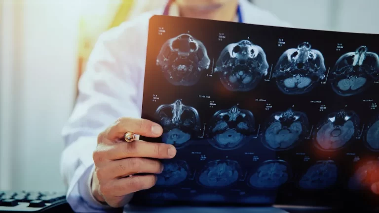
MRI (magnetic resonance imaging) radiology is a type of medical imaging that uses a powerful magnetic field, radio waves, and a computer to produce detailed images of the inside of the body. MRI scans can provide detailed images of soft tissues and organs and are commonly used to diagnose and monitor a variety of medical conditions.
During an MRI scan, the patient lies on a table that slides into a large, cylindrical machine. The machine generates a powerful magnetic field that aligns the protons in the body’s tissues. Radio waves are then sent through the body, causing the protons to produce faint signals that are detected by the machine. A computer then uses these signals to create detailed images of the body’s internal structures.
MRI radiology is a useful diagnostic tool for a variety of medical conditions, including brain and spinal cord injuries, tumors, and joint injuries. It can also be used to visualize blood vessels, diagnose heart conditions, and monitor the progression of certain diseases.
While MRI radiology is generally considered safe, it can be uncomfortable for some patients due to the enclosed space of the MRI machine. Patients who are claustrophobic may require medication or sedation to help them relax during the procedure. In addition, patients with certain medical devices, such as pacemakers, may not be able to undergo an MRI scan due to potential safety risks.
Ultrasound Radiology
Ultrasound radiology is a type of medical imaging that uses high-frequency sound waves to create images of the inside of the body. Ultrasound scans can provide real-time images of organs, tissues, and blood flow, and are commonly used to diagnose and monitor a variety of medical conditions.
During an ultrasound scan, a small handheld device called a transducer is placed on the skin over the area being imaged. The transducer emits high-frequency sound waves that bounce off the body’s internal structures and are detected by the transducer. A computer then uses these sound waves to create real-time images of the body’s internal structures on a monitor.
Ultrasound radiology is a useful diagnostic tool for a variety of medical conditions, including pregnancy, gallbladder, and liver disease, and conditions affecting the kidneys, bladder, and other organs. It is also commonly used to guide medical procedures, such as biopsies and fluid drainage.
Ultrasound radiology is generally considered safe, as it does not expose patients to ionizing radiation like X-rays or CT scans. However, it may not be suitable for all patients, particularly those who are obese or have a lot of gas in their intestines, as this can make it difficult to obtain clear images.
Nuclear Medicine Radiology
Nuclear medicine radiology is a medical specialty that uses small amounts of radioactive materials, known as radiopharmaceuticals, to diagnose and treat a variety of medical conditions. Nuclear medicine radiology in Chennai is unique in that it provides information about both the structure and function of organs and tissues.
During a nuclear medicine scan, a small amount of radiopharmaceutical is injected into the patient’s bloodstream, swallowed, or inhaled. The radiopharmaceutical then travels through the body and emits gamma rays, which are detected by a special camera. The camera produces images that show the distribution of the radiopharmaceutical within the body and provide information about organ function.
Nuclear medicine radiology in Chennai is used to diagnose and treat a wide range of medical conditions, including cancer, heart disease, and neurological disorders. It can also be used to evaluate organ function and blood flow and to monitor the effectiveness of certain treatments.
While nuclear medicine radiology uses small amounts of radiation, the radiation exposure is generally considered safe and the benefits of the test often outweigh the risks. However, patients who are pregnant or may be pregnant should inform their healthcare provider before undergoing a nuclear medicine scan, as radiation exposure can be harmful to developing fetuses.
What are the Benefits and Risks of Radiology?
Radiology is a safe and effective diagnostic tool that has many benefits over other diagnostic procedures such as surgery. It is non-invasive, painless, and does not require anesthesia. Radiology in Chennai can provide detailed images of the internal structures of the body, which can help healthcare professionals diagnose and treat various medical conditions.
However, there are some risks associated with radiology in Chennai, such as exposure to radiation. The amount of radiation exposure depends on the type of radiology being used and the area of the body being imaged. Pregnant women and children are more susceptible to the harmful effects of radiation and may need to take special precautions before undergoing radiology in Chennai.
Conclusion
Radiology in Chennai is a safe and effective diagnostic tool that is used in many different medical specialties to diagnose and treat various medical conditions. It involves the use of different types of imaging modalities such as X-rays, CT scans, MRI scans, and ultrasound to visualize the internal structures of the body. If you have any questions or concerns about radiology, please consult your healthcare professional.
Read also Diabetic Foot Clinic in Chennai.